Diagnosis encompasses:
- Clinical assessment
- Dermoscopy
- Digital mole analysis and monitoring
- Total body photography
- Biopsy
Clinical Assessment
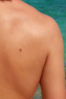
Clinical assessment with a large magnifying lamp is used to locate any spots of interest, including moles, melanoma, and non-melanoma skin cancers.
Dermoscopy
Dermoscopy eliminates the surface reflection from the skin so that subsurface features can be seen. These features would otherwise not be visible - regardless of magnification. More...
Digital Dermoscopy
Digital dermoscopic images of moles can be stored using the MoleMax system, and so can be monitored for future change. More...
Total Body Photography
Total body photography is an extra service for higher-risk patients whereby a set of images that cover the entire skin surface is made available to the patient to facilitate monitoring. More...


